Astrocytes
FCIV: Expression of reporter gene in astrocytes transduced with lentiviral vector under ubiquitin-c promoter
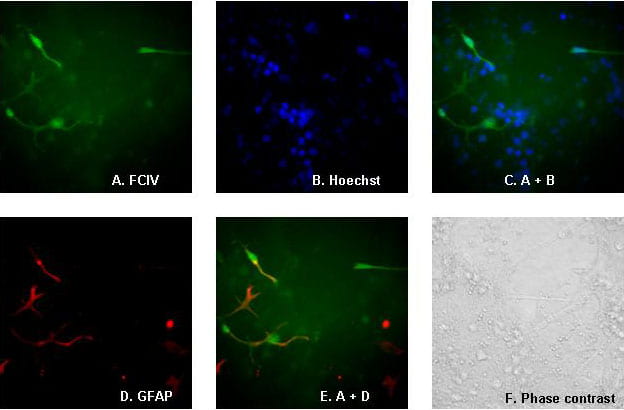
Mixed murine neocortical cultures containing both neurons and astrocytes were transduced with lentiviral vector FCIV at an MOI of 5. Images were obtained 3 days after transduction. Cultures were stained with Hoescht 33342 to visualize nuclei and immunostained with anti-GFAP antibody (rabbit polyclonal, IncStar) with Alexa 594 secondary antibody. All images are of the same field. A. GFP fluorescence. B. Hoescht 33342. C. Overlay of Hoescht staining and GFP signal. D. GFAP immunostaining. E. Overlay of GFP and GFAP immunostaining. F. Phase-contrast image. Original magnification 400X.
Expression of reporter gene in astrocytes transduced with lentiviral vector with CMV promoter
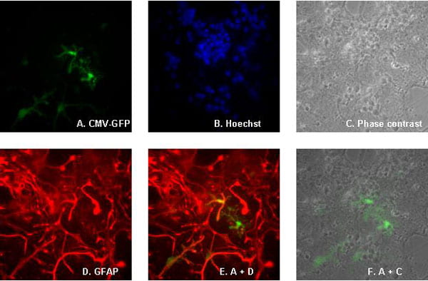
Mixed murine neocortical cultures containing both neurons and astrocytes were transduced with lentiviral vector CMV-GFP at an MOI of 5. Cultures were imaged 3 days after transduction. Cultures were stained with Hoescht 33342 to visualize nuclei and immunostained with anti-GFAP antibody (rabbit polyclonal, IncStar) with Alexa 594 secondary antibody. All images are of the same field. A. GFP fluorescence. B. Hoescht 33342. C. Phase contrast. D. GFAP immunostaining. E. Overlay of GFP and GFAP immunostaining. F. Overlay of GFP and phase-contrast image. Original magnification 400X.
Expression of reporter gene in astrocytes transduced with lentiviral vector under phosphoglycerate kinase (PGK) promoter
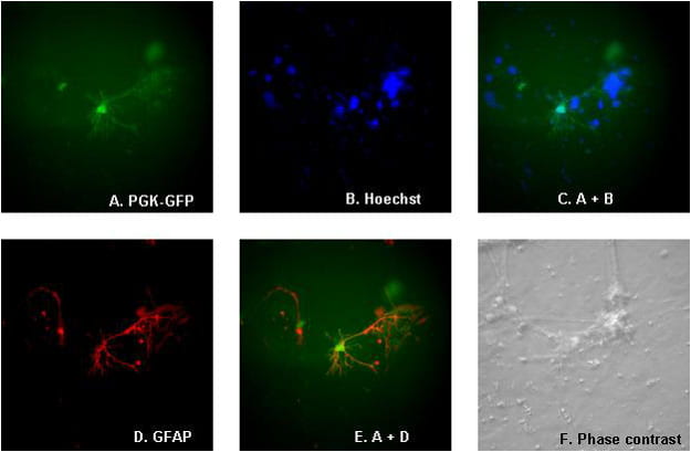
Mixed murine neocortical cultures containing both neurons and astrocytes were transduced with lentiviral vector PGK-GFP at an MOI of 5. Images were obtained 3 days after transduction. Cultures were stained with Hoescht 33342 to visualize nuclei and immunostained with anti-GFAP antibody (rabbit polyclonal, IncStar) with Alexa 594 secondary antibody. All images are of the same field. A. GFP fluorescence. B. Hoescht 33342. C. Overlay of Hoescht staining and GFP signal. D. GFAP immunostaining. E. Overlay of GFP and GFAP immunostaining. F. Phase-contrast image. Original magnification 400X.
Transgene expression in neocortical cultures transduced with lentiviral vectors under GFAP promoter
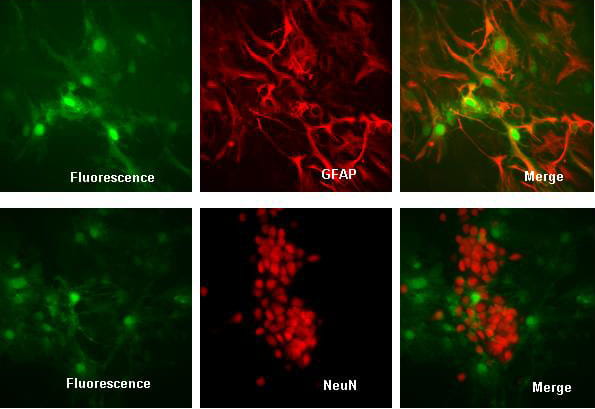
Glial fibrillary acidic protein (GFAP) promoter in the vector drives expression of green fluorescent protein (GFP) only in astrocytes as verified by colocalization with GFAP but not NeuN. Primary murine neocortical cultures containing both neurons and astrocytes were transduced with the lentiviral vector GFAP-GFP at an MOI of 5. Seven days after transduction, cultures were fixed and immunostained with anti-GFAP (rabbit polyclonal antibody, IncStar, Alexa 594 secondary antibody) shown in upper 3 panels, or with anti-NeuN (Chemicon antibody, Alexa 594 secondary) shown in lower 3 panels. Left panels show GFP fluorescence, middle panel shows Alexa 594 signal, right panel shows overlay of GFP and immunostaining. Original magnification was 400X. Note GFP signal localizes to both nuclei and processes, while GFAP staining spares nuclei.
Expression of reporter gene in neurons transduced with lentiviral vector under myeloproliferative sarcoma virus enhancer (MND) promoter
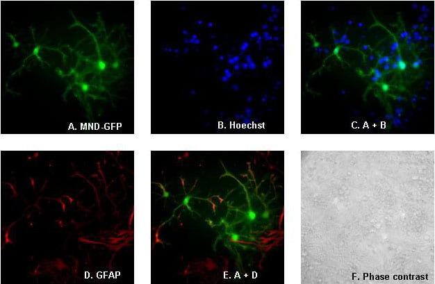
Mixed murine neocortical cultures containing both neurons and astrocytes were transduced with lentiviral vector MND-GFP at an MOI of 5. Images were obtained 14 days after transduction. Left panel: GFP expression. Right panel: phase contrast image. Original magnification 400X.