Neurons
Expression of reporter gene in neurons transduced with lentiviral vector under ubiquitin-c promoter
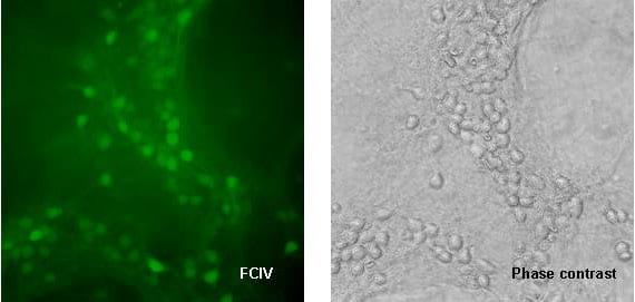
Mixed murine neocortical cultures containing both neurons and astrocytes were transduced with lentiviral vector FCIV at an MOI of 5. Images were obtained 14 days after transduction. Left panel: YFP venus expression. Right panel: phase contrast image. Original magnification 400X.
Expression of reporter gene in neurons transduced with lentiviral vector under phosphoglycerate kinase (PGK) promoter
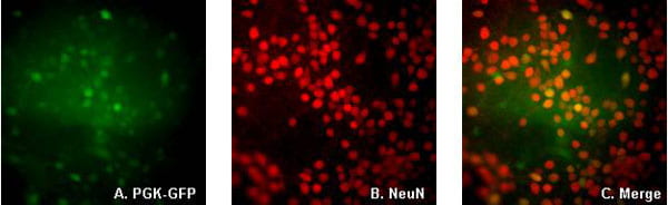
Mixed murine neocortical cultures containing both neurons and astrocytes were transduced with lentiviral vector PGK-GFP at an MOI of 5. Cultures were immunostained and imaged 7 days after transduction. Cultures were immunostained with NeuN (chemicon) with Alexa 594 secondary antibody. A. GFP fluorescence, B. NeuN immunocytochemistry, C. Overlay of GFP and NeuN. Original magnification 400X.
Comparison of neurons transduced with PGK-Lck-GFP and PGK-GFP
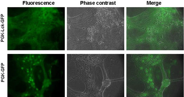
Murine neocortical cultures containing both neurons and astrocytes were transduced with lentiviral vector PGK-GFP or PGK-lck-GFP, in which a membrane-targeting Lck sequence is fused to N terminus of eGFP. In contrast with the original eGFP that primarily expressed in neuron cell bodies, the Lck-GFP fusion protein was almost exclusively localized in processes. Left panel shows GFP fluorescence, middle panel is phase contrast image of the same field and right panel shows overlay of phase contrast and GFP images. Original magnification 400X.
Expression of reporter gene in neurons transduced with lentiviral vector under myeloproliferative sarcoma virus enhancer (MND) promoter
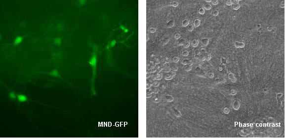
Mixed murine neocortical cultures containing both neurons and astrocytes were transduced with lentiviral vector MND-GFP at an MOI of 5. Images were obtained 14 days after transduction. Left panel: GFP expression. Right panel: phase contrast image. Original magnification 400X.5
Transgene expression in neocortical cultures transduced with lentiviral vectors under synapsin promoter
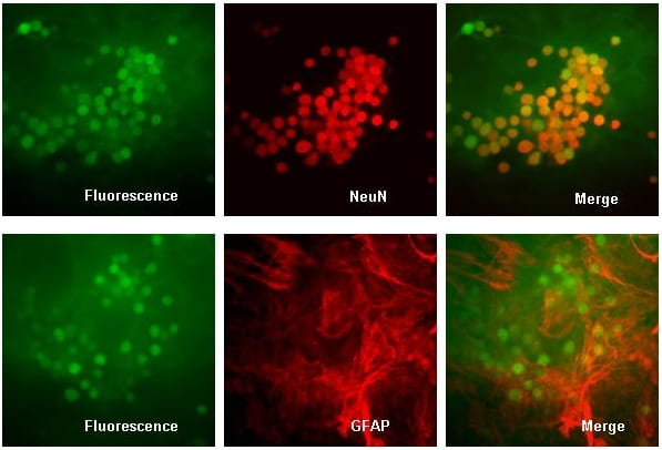
Synapsin (SYN) promoter in the vector drives expression of green fluorescent protein (GFP) only in neurons as verified by colocalization with NeuN but not GFAP. Primary murine neocortical cultures containing both neurons and astrocytes were transduced with the lentiviral vector SYN-GFP at an MOI of 5. Seven days after transduction, cultures were fixed and immunostained with anti-NeuN antibody (Chemicon, Alexa 594 secondary antibody) shown in upper 3 panels, or with anti-GFAP antibody (rabbit polyclonal antibody, IncStar, Alexa 594 secondary antibody) shown in lower 3 panels. Left panels show GFP fluorescence, middle panel shows Alexa 594 signal, right panel shows overlay of GFP and immunostaining. Original magnification was 400X.