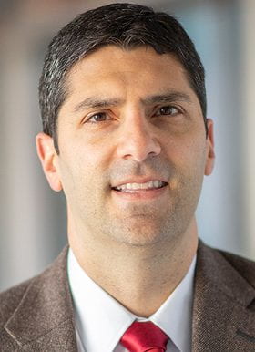
Brian Gordon, PhD
Assistant Professor, WashU Radiology
- Phone: 314-747-7354
- Email: bagordon@nospam.wustl.edu
Using in vivo neuroimaging and biofluid measurements in humans to better understand the pathophysiology of Alzheimer disease
The mission of the Hope Center is to improve the lives of individuals with neurological disorders through collaborative, and translational research. These goals are core tenants of my own work. My research focuses on using in vivo neuroimaging and biofluid measurements in humans to better understand the pathophysiology of Alzheimer disease (AD). The primary neuroimaging techniques that I utilize are magnetic resonance imaging (MRI) and positron emission tomography (PET). Additionally, I have incorporated cerebrospinal fluid (CSF) measures such as total tau (t-tau), phosphorylated tau (p-tau181), beta-amyloid (Ab), and neurofilament light chain (NFL) into my work. The primary goal of my research is two-fold. The first main area of academic interest focuses on characterizing the temporal progression and evolution of the different markers of amyloidosis and neurodegeneration that we can measure in vivo. I study this question in both autosomal dominant and sporadic forms of AD. By understanding the temporal relationships of the biomarkers, as well as their interrelationships, we can formulate theories about the cascade of biological changes that eventually result in dementia. The second focus is on using what we know about these biomarkers to aid clinicians. This includes separating dementias with overlapping phenotypes but divergent etiologies (e.g. AD and frontotemporal dementia), identifying atypical subtypes of AD (e.g. visuospatial or language predominant AD), as well as assessing what factors predict the onset of AD dementia in otherwise cognitively normal older adults.
More about the Gordon Neuroimaging Lab